Search
Close this search box.

Transesophageal Echocardiogram (TEE) Patient Information

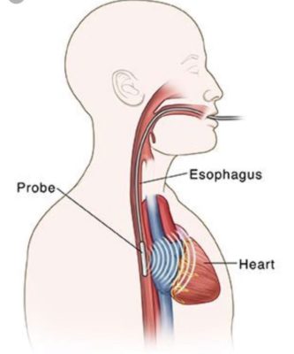
A Transesophageal Echocardiogram (TEE) is a diagnostic test that provides a clearer view of the heart compared to a regular transthoracic echocardiogram (TTE). This is because the probe used in TEE is placed in the esophagus, which lies close to the heart, avoiding interference from the ribs or lungs. The TEE probe also has 3D imaging capabilities, allowing for highly detailed images of the heart’s structures.


Common Indications for TEE

  • Valve Assessment: Evaluation of valve structure, including the degree of narrowing (stenosis) or leakage (regurgitation).
  • Clots: Checking for the presence of cardiac thrombus (blood clots), especially in the context of stroke.
  • Heart Defects: Diagnosing abnormal openings between heart chambers, such as a patent foramen ovale (PFO) or atrial septal defect (ASD), which can cause strokes or heart failure.
  • Inconclusive TTE: When a regular echocardiogram provides suboptimal results, TEE offers clearer images (e.g., in patients with thick chest walls or obesity).
  • Valvular Infections: Diagnosis of infections affecting the heart valves, such as endocarditis, or detecting complications of the infection.
  • Post-Surgery Follow-Up: Monitoring the condition of surgically repaired heart valves.
  • Congenital Heart Conditions: Follow-up for individuals with congenital heart disease.

Duration: The procedure itself lasts around 30 minutes, but you will spend approximately 4 hours at the hospital, including preparation and post-procedure recovery.

Procedure Steps:

  • Preparation: A nurse will place an intravenous (IV) line in your arm and administer a sedative to help you relax. Your throat will be sprayed with a numbing agent to suppress the gag reflex.
  • Monitoring: During the procedure, your vital signs will be monitored through an electrocardiogram (EKG) and a pulse oximeter (measuring your oxygen levels).
  • Probe Insertion: The cardiologist will gently guide a flexible, thin tube (probe) through your mouth and down your throat. You will be asked to swallow as the probe advances into the esophagus.
  • Imaging: The transducer at the end of the probe emits sound waves to capture detailed pictures of your heart, which are displayed on a video screen.
  • Completion: After the images are taken, the probe, IV, and monitoring electrodes will be removed. You will remain under observation until fully awake. Due to sedation, you must arrange for someone to drive you home.


Preparation Before TEE:

  • Fasting: If your test is scheduled in the morning, do not eat or drink anything after midnight. For afternoon appointments, fast for at least 6 hours before the procedure.
  • Medications: If instructed to take medications before the test, take them with small sips of water. Do not stop taking blood thinners unless directed by your doctor.
  • Diabetes Management: If you take insulin or oral diabetes medications, skip your morning dose if you have not eaten for at least 6 hours before the procedure.
  • Hospital Call: Expect a call from the hospital the evening before the procedure with specific instructions, including the time, location, and address.


After TEE:

  • Numb Throat: Your throat may be numb for a short period, so do not eat or drink for at least 4 hours after the procedure to avoid choking.
  • Sore Throat: You may experience mild throat soreness for a day or two.
  • Driving: Because of sedation, do not drive for the rest of the day. You may resume normal activities the following day.
  • Results: You will receive the results of the test immediately after the procedure, but the sedatives may affect your memory, so the person driving you home can help recall the information.

The TEE offers highly detailed, 3D images of your heart, helping your cardiologist make more accurate diagnoses and treatment plans.